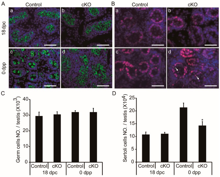
Figure 4.
Accumulated tubular germ cells and decreased Sertoli cells in neonatal Ddb1 cKO mice. (A) Testicular sections show germ cells (DDX4, green) in 18 dpc and 0 dpp mice, scale bars = 50 μm; (B) testicular sections show Sertoli cells (SOX9, red) in 18 dpc and 0 dpp mice, arrows indicate Sertoli cells that aberrantly located in the lumen of testis cords, scale bars = 50 μm; (C) the total number of germ cells per testis shown in (A); and (D) the total number of Sertoli cells in testicular section per testis as shown in (B). Data were presented as mean ± S.E.M. * p < 0.05, Student’s t-test.

