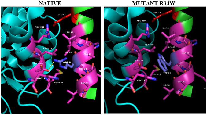
Figure 4.
The PAN-associated novel mutation R34W in the ADA2 structure. Panels illustrate the 3D native structure of ADA2 (PDB 3LGG) and mutant on position #8. A section of the ADA2 dimer interface is shown, illustrating the residue contacts between the two HN1 helix anchors, where Arg34 (blue-grey) is located. The Arg34Trp mutant causes severe clashes between the bulky side chain of the tryptophane 34 side chain and Leu372 of the homodimer's a5 helix as well as loss of the dimer stabilizing hydrogen bond interaction (in yellow dashed lines) between Arg34 (blue-grey) and homomonomer Asp373 (red). Structures were analyzed, mutants constructed and cartoons rendered using the PyMOL Molecular Graphics System, Version 2.2. ADA2, adenosine deaminase 2; PAN, polyarteritis nodosa.

