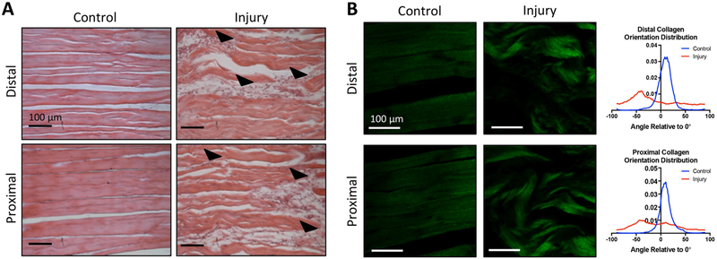
Figure 1: One week after injury tendon undergoes noticeable damage.
(A) H&E staining of longitudinally-sectioned tendon reveals damage 1 week after injury, indicated by changes in collagen structure and cellular infiltration (arrowheads) (B) Second Harmonic Generation images of collagen within tendon sections with quantification of orientation distribution. The average alignment of collagen fibers in the control supraspinatus tendon relative to 0° was 8.5° ± 16.7° (distal) and 9.9° ± 16.0° (proximal), while the injured supraspinatus tendon had an average collagen alignment of −11.8° ± 51.7° (distal) and −8.3° ± 46.9° (proximal) relative to 0°. The orientation of collagen fibers in injured supraspinatus tendon was significantly different from control (p<0.05). Scale bars are 100 μm.

