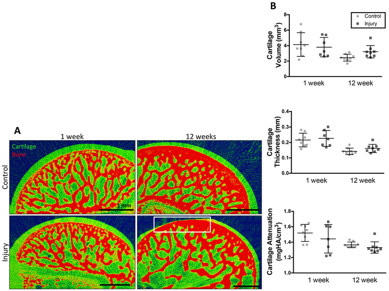
Figure 3: Contrast enhanced microCT reveals focal defects within cartilage 12 weeks post-injury.
(A) contrast enhanced microCT of humeral heads. Loss of cartilage (focal defects) highlighted in white box. Defects present in 4 out of 8 animals. N=8. scale bar = 1mm. (B) Evaluation of cartilage thickness, cartilage volume, and cartilage attenuation. N=8, no significant differences present.

