Fig. 6.
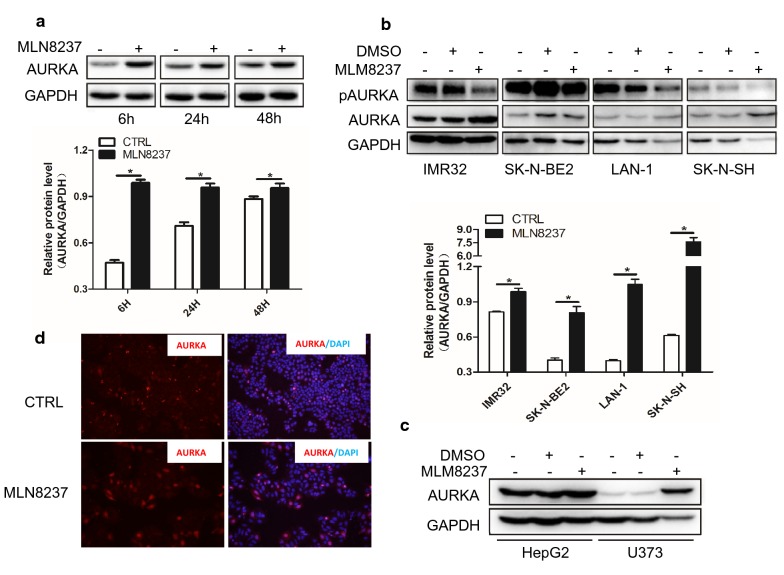
MLN8237 treatment led to abnormal high expression of AURKA in vitro. a IMR32 cells were treated with 2 μmol/l of MLN8237. Cell samples were collected at 6 h, 24 h, and 48 h time points. Total proteins were extracted for western blot analysis for AURKA protein expression. b Different neuroblastoma cell lines including IMR32, SK-N-BE2, LAN-1, and SK-N-SH were treated with 2 μmol/l of MLN8237. At 48 h, total proteins were extracted for western blot to test AURKA and pAURKA expression. Gray values were calculated by imageJ software. The data were shown as the mean ± SEM of three independent experiments. *P < 0.05. c Hepatocellular carcinoma cell line HepG2 and glioma cell line U373 were treated with 2 μmol/l of MLN8237. At 48 h, total proteins were extracted for western blot to test AURKA and pAURKA expression. d 1.0 × 10⁵ of IMR32 cells were seed into 8-well chamber slide. The next day, cells were treated with 2 μmol/l of MLN8237. At forty-eight hours after transfection, cells were fixed and Immunofluorescence staining was performed to test AURKA expression

