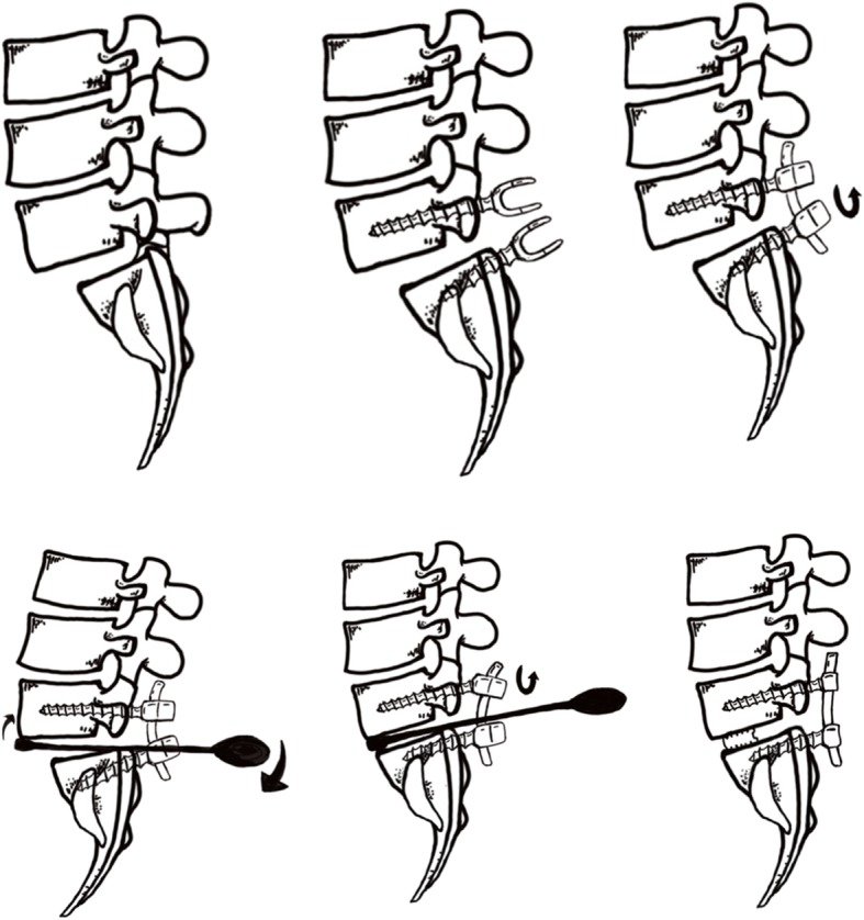
Fig. 2.

Reduction process of a slipped vertebrae. a, Forward slippage of L5; b, Pedicle screws were placed at both vertebra of the slipped levels; c, The nerve roots were decompressed before reduction. After removal of the disk tissues and endplate preparation, a rod was placed unilaterally and the pedicle screw of the lower vertebrae was locked; d, A lever repositioner was placed at the anterior rim of the slipped vertebrae under fluoroscopy; e, With the lower vertebrae as the lever fulcrum, force was applied to gradually pry the slipped vertebrae upward; f, The pedicle screws of the slipped vertebrae were locked. Then, an addition rod was placed and all screws were locked. The extent of slip reduction was verified with fluoroscopy. After reduction, the interspace was packed with autologous bone graft material and an appropriate cage filled with bone was inserted into the disc space
