Figure 2.

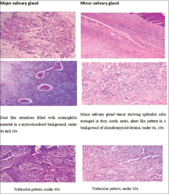
Photomicrographs of the hematoxylin and eosin-stained tissue sections showing cellular components, stromal components, capsular alterations and inflammatory changes of pleomorphic adenoma of major and minor salivary glands

Photomicrographs of the hematoxylin and eosin-stained tissue sections showing cellular components, stromal components, capsular alterations and inflammatory changes of pleomorphic adenoma of major and minor salivary glands