Figure 3.

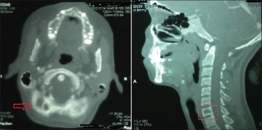
Photograph of computed tomography image of neck area showing a radiolucent lesion at C7 level both in coronal (show by arrow) and lateral (indicated in the box) views

Photograph of computed tomography image of neck area showing a radiolucent lesion at C7 level both in coronal (show by arrow) and lateral (indicated in the box) views