Figure 8.

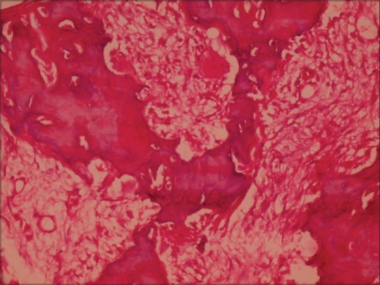
Photomicrograph of hematoxylin and eosin stained decalcified sections show multinucleated giant cells and plump cementoblasts (H&E stain, ×40)

Photomicrograph of hematoxylin and eosin stained decalcified sections show multinucleated giant cells and plump cementoblasts (H&E stain, ×40)