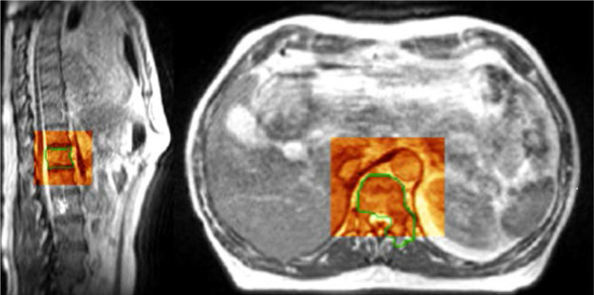
Figure 2.
Example slices (sagittal and axial) from 3D setup volume at time of treatment for Co-60 MRI-RT SABR of a L1 spinal metastasis. The grayscale image represents the MRI simulation image. The red shaded box demonstrates the aligned setup imaging of the day. A green outline delineates the PTV which includes the vertebral body and left pedicle. The bright cerebral spinal fluid, spinal cord, and bony anatomy are clearly visualized in the spinal canal on the sagittal image. On the axial image, the tip of the spinal cord and cauda equina are observed posteriorly in the dural sac. 3D,three-dimensional; SABR, stereotactic ablativeradiotherapy; PTV, planning target volume.

