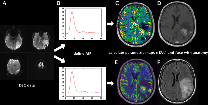
Figure 1. .
Workflow of highly standardized preprocessing of raw DSC-perfusion data sets (A). After background segmentation, removal of extracranial tissue and noise threshold detection, the raw signal was converted into relative change in R2* vs time and the AIF was generated automatically (B). We used the same presets for both software suites and calculated hemodynamic parameter maps of rBV, rBF and MTT. Example shows rBVc map fused with T1W + Gd produced with NordicICE (C) and with Olea sphere (E). Anatomical images T1W + Gd (D) and T2W FLAIR (F) of a representative brain tumor case (left parietooccipital glioblastoma). AIF, arterial inputfunction; DSC, dynamic susceptibility contrast; FLAIR, fluidattenuated inversion recovery; MTT, mean transit time; rBF, relative cerebralblood flow; rBV, relative cerebral blood volume.

