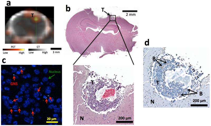
Figure 5.
Sub-millimeter brain tumor detection with 124I-GNS. (a) Sub-millimeter brain tumor identified on PET/CT image obtained 48 h post 124I-GNS injection in Mouse 3. The average tumor uptake was 0.66 %ID/g and the T/N was 4.7. (b) H&E histopathology examination confirmed the identified brain tumor region from PET/CT imaging. The identified tumor was less than 0.5 millimeter in size. (c). TPL imaging showed that the GNS (white spots, marked by red arrow) were inside the tumor. The tumor cell nuclei were stained with DAPI (blue). (d) CD31 immunohistochemical staining confirmed the presence of endothelial cells and the developing vasculature. Two blood vessels (B) parallel to the tumor section surface were identified. (T), tumor; (N) normal brain.

