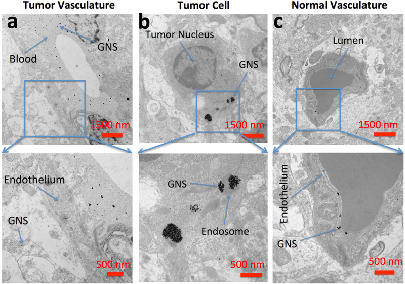
Figure 6.
Electron microscopy of brain tumor 24 hours after intravenous administration of GNS (a) TEM imaging of GNS in the extracellular space of the tumor region. (b) TEM imaging of GNS in endosomes within brain tumor cells. (c) TEM imaging of GNS in normal brain vasculature. PEGylated GNS nanoparticles leak through brain tumor vasculature, diffuse into tumor extracellular space and are endocytosed inside tumor cells. In normal brain, GNS were confined inside the vasculature wall, consistent with an intact BBB. Scale bar, 1,500 nm (top row) and 500 nm (bottom row).

