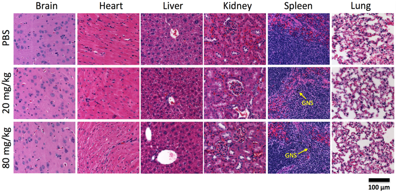
Figure 8.
Histopathology examination of peripheral organs. Histopathology following IV administration of GNS H&E histopathology examination of brain, heart, liver, kidney, spleen and lung from mice obtained 6 months after PBS or GNS injection (20 mg/kg or 80 mg/kg dose). Scale bar, 100 μm. The GNS (black color) nanoparticles were seen inside spleen for mice in both 20 mg/kg and 80 mg/kg dose groups. H&E evaluation was unremarkable and demonstrated healthy and intact tissue.

