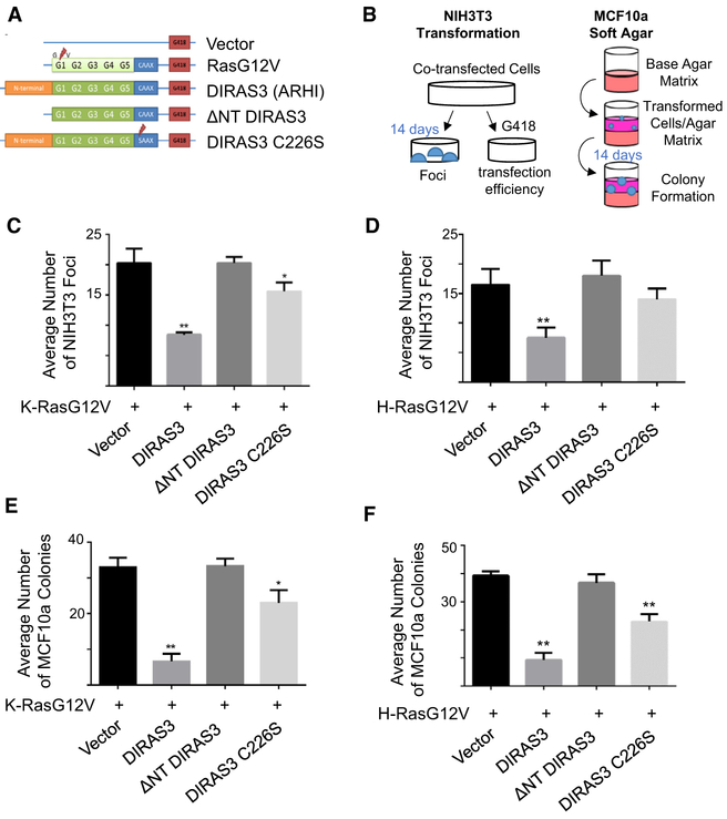
Figure 1. DIRAS3 Inhibits RAS-Induced Malignant Transformation of Murine Fibroblasts and RAS-Induced Anchorage-Independent Growth of Partially Transformed Human Breast Epithelial Cells.
(A) Illustration of constructs in pcDNA3.1 vectors that were used for the following experiments.
(B) Experimental schema for the assays performed.
(C and D) DIRAS3 inhibits K-RAS- (C) and H-RAS-induced (D) transformation of murine NIH 3T3 cells. Cells were plated in 60-mm dishes and transfected with 10 μg of each DNA plasmid for 24 h prior to being separated into dishes for focus formation and clonogenic selection by G418. Transformed foci were counted at 10× magnification as they appeared within two 10 × 10-mm areas per plate. The columns indicate the mean, and the error bars indicate the SD (**p < 0.01; *p < 0.05). Clonogenic selection by G418 was used to ensure equal transfection efficiency.
(E and F) DIRAS3 inhibits anchorage-independent growth of MCF10a breast epithelial cells transformed with K-RAS (E) and H-RAS (F). Cells were plated in 6-well plates and transfected with 3 μg of each DNA plasmid for 24 h to being re-seeded at a cell density of 1.0 × 10⁵ in soft agar. Cells were grown for 2 weeks and colonies were counted. The assay was performed three times with technical triplicates for each experiment. The columns indicate the mean, and the error bars indicate the SD (**p < 0.01).

