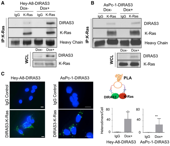
Figure 3. DIRAS3 Co-localizes with K-RAS.
(A and B) Co-immunoprecipitation of DIRAS3 and K-RAS. Hey-A8-DIRAS3- (A) and AsPc-1-DIRAS3- (B) inducible cells at 70% confluency in 60-mm dishes were treated with or without DOX for 48 h. Cell lysate was collected, and 1.5 ug of lysate was used for immunoprecipitation with a rabbit immunoglobulin G (IgG) control or anti-K-RAS antibody. Western blotting was performed and probed with anti-DIRAS3 antibody before stripping and probing with anti-K-RAS.
(C) DIRAS3 and K-RAS formed heterodimers in Hey-A8-DIRAS3 cells. DIRAS3 and K-RAS complexes were analyzed with an in situ PLA assay. Scale bars represent 20 μm. Data were obtained from two independent experiments performed in duplicate. Columns indicate the mean, and the bars indicate the SD (**p < 0.01).

