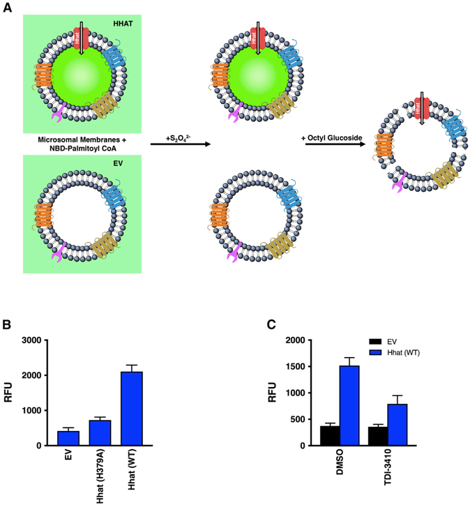
Figure 1. NBD-Palmitoyl-CoA Uptake Assay.
(A) Microsomal membranes generated from HEK293FT cells transfected with pcDNA3.1 (empty vector; EV) or pcDNA3.1 encoding Hhat are incubated with NBD-palmitoyl-CoA, then treated with dithionite (Na2S2O42−), a membrane-impermeant reducing agent that quenches NBD fluorescence of molecules on the external side of the membrane. RFU readings at 535 nm reflect NBD-palmitoyl-CoA within the vesicle and protected from the quencher. The addition of 0.2% octyl glucoside permeabilizes the bilayer and allows access of the quencher to the interior of the vesicle.
(B) Microsomal membranes from HEK293FT cells expressing pcDNA, Hhat WT, or Hhat H379A were incubated with NBD-palmitoyl-CoA for 60 min at room temperature prior to the addition of Na2S2O42−. NBD fluorescence in the interior of the vesicles was quantified; n = 3.
(C) Microsomal membranes were pretreated for 15 min with 10 μM TDI-3410, then analyzed for NBD-palmitoyl-CoA; n = 3.

