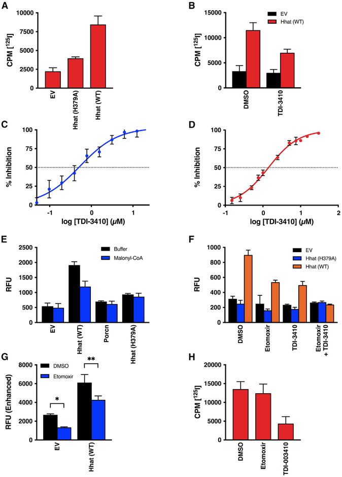
Figure 2. Hhat Enhances Uptake of Palmitoyl-CoA across the ER Membrane.
(A) Microsomal membranes from HEK293FT cells expressing pcDNA, Hhat WT, or Hhat H379A were incubated with [125I] IodoPalmitoyl-CoA for 60 min at room temperature prior to the addition of 2% BSA. 125I cpm remaining inside the vesicles after washing was quantified; n = 2.
(B) Microsomal membranes were pretreated for 15 min with 10 μM TDI-3410, then analyzed for [125I] IodoPalmitoyl-CoA uptake; n = 3.
(C and D) Microsomal membranes containing WT Hhat were pretreated for 15 min with the indicated concentrations of TDI-3410.
(C) The amount of NBD-palmitoyl-CoA remaining within the vesicle was quantified; n = 3; IC50 = 0.5 μM.
(D) Membranes were assayed for Shh palmitoylation activity by incubation with biotinylated Shh peptide and [125I] IodoPalmitoyl-CoA; n = 2; IC50 = 1.4 μM.
(E and F) Microsomal membranes from HEK293FT cells expressing Hhat or Porcupine were pretreated for 15 min with (E) 5 μM malonyl-CoA, (F) 10 μM TDI-3410, 10 μM Etomoxir, or both TDI-3410 and Etomoxir. NBD-palmitoyl-CoA uptake was determined (n = 3) as in Figure 1.
(G) As in (F), but with the gain on the plate reader increased; n = 3, *p = 0.03; **p = 0.003.
(H) Purified Hhat was incubated with 10 μM TDI-3410 or 10 μM Etomoxir, and Shh palmitoylation activity was assayed; n = 3.
See also Figure S1.

