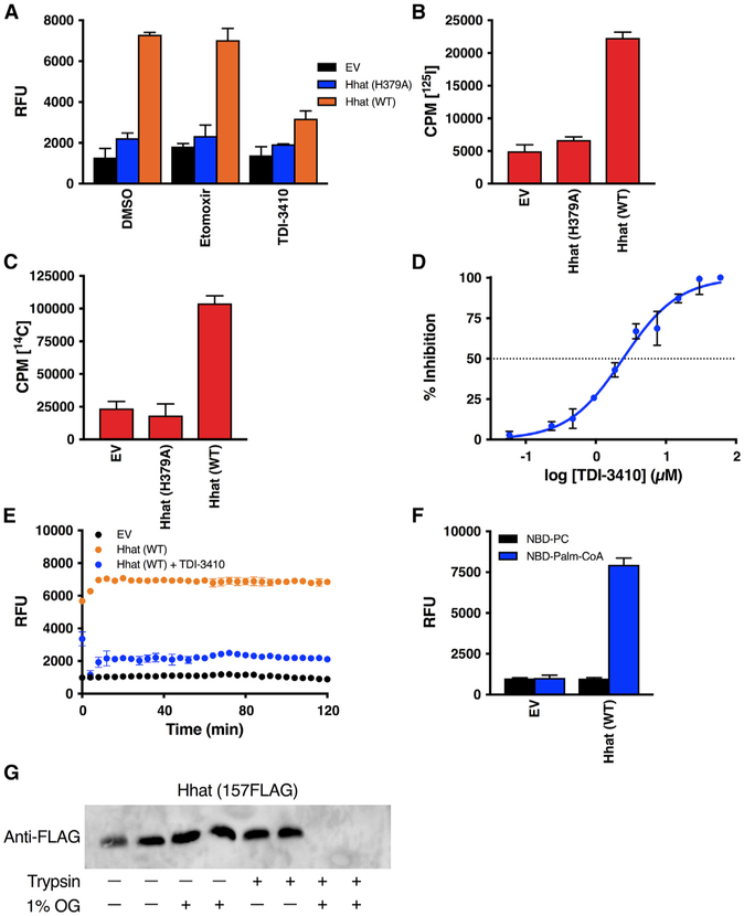
Figure 4. Purified Hhat Reconstituted into Artificial Liposomes Mediates Palmitoyl-CoA Uptake.
(A–C) The 0.2-μm liposomes reconstituted with or without purified Hhat WT or Hhat H379A were pretreated for 15 min with 10 μM TDI-3410 or 10 μM Etomoxir (A) and incubated for 60 min at room temperature with NBD-palmitoyl-CoA (A; n = 4), [125I] IodoPalmitoyl-CoA (B; n = 2), or [14C] palmitoyl-CoA (C; n = 2), and palmitoyl-CoA uptake was determined as in Figures 1 and 2.
(D) Liposomes reconstituted with purified Hhat WT were pretreated with the indicated concentrations of TDI-3410 and NBD-palmitoyl-CoA uptake was analyzed as in (A); n = 3.
(E) As in (A). After quencher addition, RFU readings were taken every 2 min for 120 min; n = 3.
(F) As in (A). Liposomes were incubated with either NBD-PC or NBD-palmitoyl-CoA; n = 2.
(G) Liposomes reconstituted with purified 157FLAG Hhat were treated with or without trypsin or octyl glucoside (OG), and Hhat was analyzed by SDS-PAGE and western blotting with anti-FLAG antibody.

