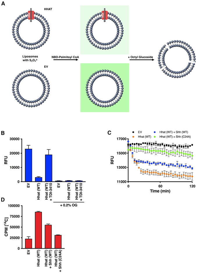
Figure 5. Quenching of Internalized NBD Fluorescence in Liposomes Containing Dithionite.
(A) Diagram of the modified NBD-palmitoyl-CoA uptake assay. Liposomes were reconstituted with or without purified Hhat (WT or H379A) in the presence of Na2S2O42−. Liposomes were re-isolated and then incubated with NBD-palmitoyl-CoA, and NBD fluorescence was quantified. The addition of 0.2% octyl glucoside reduced fluorescence levels to background.
(B) Liposomes generated as in (A) were pretreated with 10 μM TDI-3410 for 15 min followed by incubation with NBD-palmitoyl-CoA for 60 min. RFU readings were taken before and after addition of 0.2% octyl glucoside; n = 3.
(C) Liposomes were reconstituted with purified WT Hhat, Na2S2O42−, and WT or C24A Shh peptide. After the removal of the external quencher and peptide, NBD-palmitoyl-CoA was added, and the fluorescent signal was monitored for 120 min; n = 3.
(D) Liposomes were reconstituted with purified WT Hhat, and uptake of [14C] palmitoyl-CoA was measured as in Figure 4C; n = 2.

