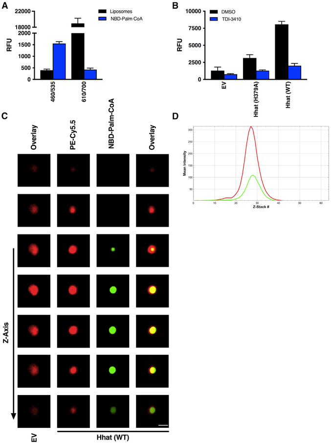
Figure 6. Visualization of Palmitoyl-CoA Uptake by Liposomes Containing Purified, Reconstituted Hhat.
(A) RFU readings at 460/535 nm and 610/700 nm. Fluorescent intensity of liposomes containing Cy5.5-PE integrated into the membrane or NBD-palmitoyl-CoA alone at 460/535 nm and 610/700 nm was determined; n = 3. No signal inference between the signals for the Cy5.5 and NBD was observed.
(B and C) Membrane-integrated Cy5.5-PE liposomes reconstituted with or without purified Hhat (WT or H379A) were pretreated with 10 μM TDI-3410 for 15 min followed by incubation with NBD-palmitoyl-CoA for 60 min at room temperature prior to quenching with Na2S2O42−.
(B) RFU readings; n = 3.
(C) Images of liposomes containing membrane integrated PE (red: Cy5.5-PE) and palmitoyl-CoA (green: NBD-palmitoyl-CoA) were acquired by confocal microscopy. Scale bar, 0.2 μm.
(D) Line scan of fluorescence intensities of Z stacks from Video S1.

