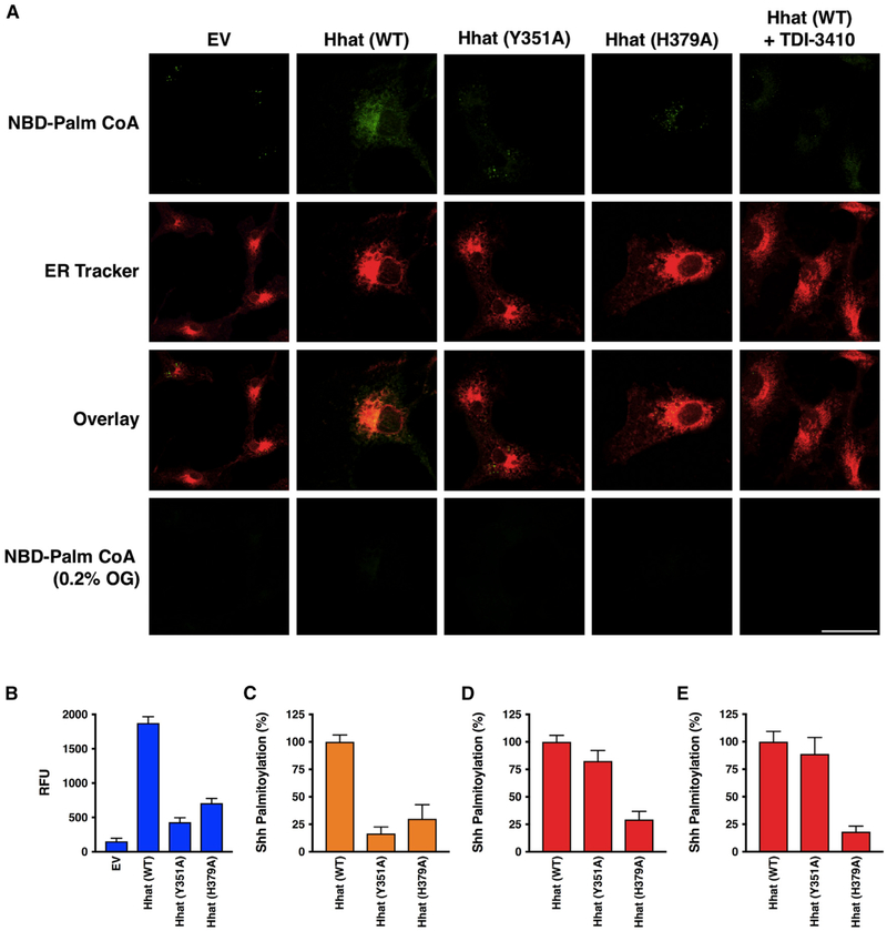
Figure 7. Palmitoyl-CoA Uptake and Shh Palmitoylation in Cells Expressing Hhat.
(A) COS-7 cells expressing EV, Hhat WT, Hhat H379A, or Hhat Y351A were incubated on ice with 65 μg/ml digitonin for 10 min to permeabilize the plasma membrane, then incubated for 60 min at room temperature with 8 μM NBD-palmitoyl-CoA. 30 min prior to imaging, 1 μM ER Tracker Red was added to the cells. Cells were washed and then imaged by confocal microscopy. Scale bar, 25 μm.
(B) NBD-palmitoyl-CoA uptake was monitored in microsomal membranes from HEK293FT cells expressing EV, Hhat WT, Hhat H379A, or Hhat Y351A as described in Figure 2; n = 3.
(C) Shh palmitoylation was monitored in COS-1 cells expressing Shh and either pcDNA, WT, or mutant (H379A, Y351A) Hhat. Cells were incubated with 15 μCi [125I] IodoPalmitate for 4 h, lysed, and Shh immunoprecipitated from cell lysates was analyzed by SDS-PAGE. [125I] CPM incorporated into Shh was quantified by phosphorimaging; n = 2.
(D) Shh palmitoylation was monitored in microsomal membranes from (B). Membranes were incubated with biotinylated Shh peptide and [125I] IodoPalmitoyl-CoA for 60 min prior to the addition of streptavidin-agarose beads. Beads were washed, and [125I] CPM incorporated into the Shh peptide were determined in a gamma counter; n = 2. See also Figure S2.
(E) Shh palmitoylation activity was assayed using purified Hhat as in (D); n = 2.

