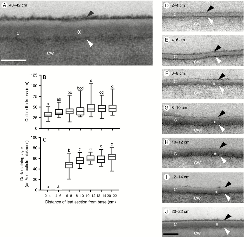
Fig. 5.
B73 leaf pavement cell cuticle development visualized by TEM. (A) Pavement cell cuticle from a partially expanded leaf 8, 40–42 cm from the base, where leaf tissue is mature. Four distinct layers or zones are visible: a thin, darkly stained layer (white arrowhead) at the interface between the cell wall (CW) and cuticle, dark (asterisk) and light zones of the cuticle proper (C), and a darkly stained epicuticular layer (black arrowhead). Scale bar = 40 nm. (B) Thickness of pavement cell cuticles at the indicated positions along the developmental gradient of partially expanded B73 leaf 8. (C) Percentage of cuticle thickness at indicated positions occupied by the dark-staining inner layer of the cuticle proper. In (B) and (C), lower-case letters indicate significance groups identified by one-way ANOVA with the Tukey multiple comparisons post-test. (D–J) Representative images of pavement cell cuticles at the indicated positions along the developmental gradient of partially expanded B73 leaf 8. Scale bar = 100 nm.

