Figure 6.
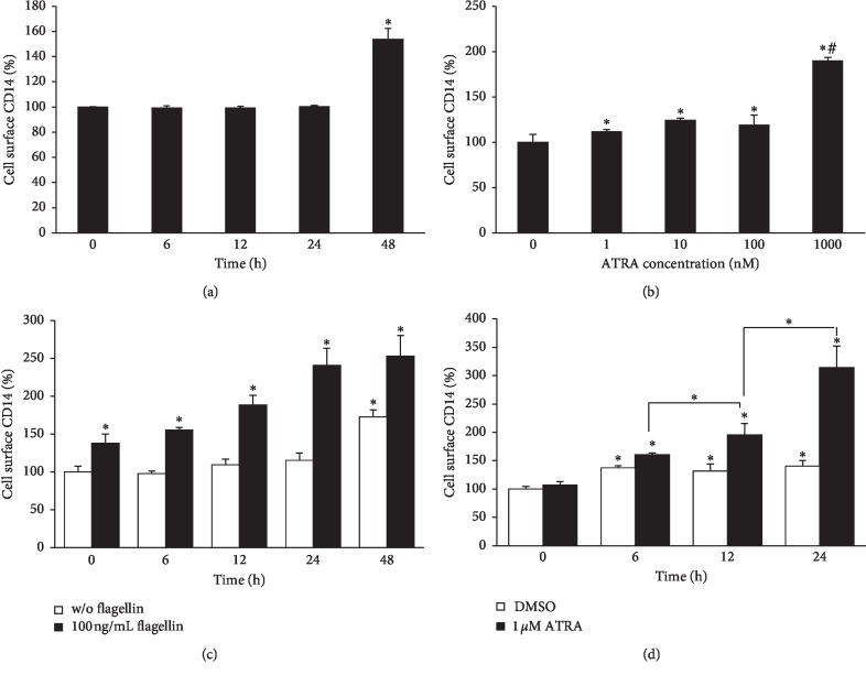
ATRA enhances the cell surface CD14 expression in THP-1 cells. Cells were treated with 1 μM ATRA for different time points (a) or with different concentrations of ATRA for 48 h (b). Cells were treated with 1 μM ATRA for different time periods as indicated and followed by challenge with 100 ng/ml flagellin from S. typhimurium for 24 h (c). Cells were treated with 1 μM ATRA for 24 h and then subjected to 100 ng/ml flagellin from S. typhimurium for different time periods as indicated (d). Cell surface expression of CD14 was measured by flow cytometry. Bar graphs indicate relative fluorescence intensity ± SD. Statistical significance was assessed by one-way ANOVA using SPSS. ∗p < 0.05 vs. DMSO control; #p < 0.05 vs. 1–100 nM ATRA-treated groups (b).

