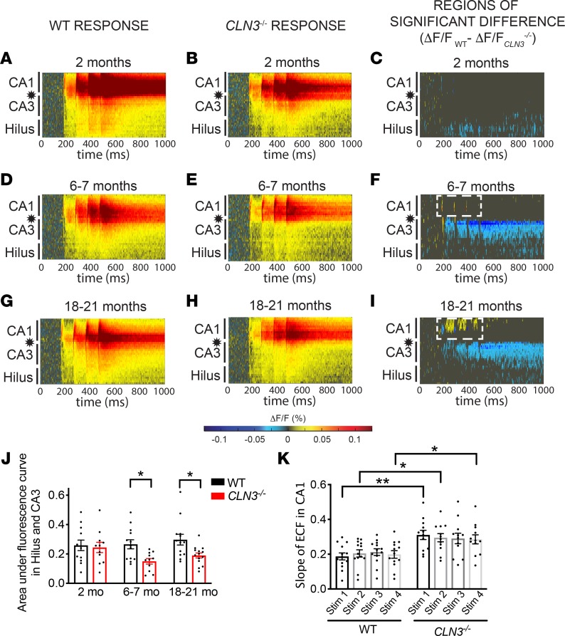
Figure 3. As CLN3 disease progresses, CA1 becomes hyperexcitable in response to SC stimulation.
(A and B) Raster plots of VSDI signal after SC stimulation (location indicated by star) reveals robust excitation of the stratum radiatum of the CA proper in 2-month-old WT and CLN3–/– hippocampal slices. (C) Permutation sampling analysis shows hypoexcitability of the hilus after SC stimulation at 2 months of age. (D–F) By 6–7 months, the CLN3–/– hippocampus demonstrates hypoexcitability of the hilus and CA3 regions as compared with WT. However, in CA1 there is emerging increased peak fluorescence in response to SC stimulation (area of warm-colored peaks, demarcated by dashed, white line in F). (G–I) By late-stage disease (18–21 months) the CA1 region of the CLN3–/– hippocampus is hyperexcitable after SC stimulation as compared with WT (region of increased activation outlined by white, dashed line). (J) Quantification of total fluorescence change in the hilus and CA3 regions confirms hypoexcitability in CLN3–/– mice at 6 and 18 months (2-way ANOVA, followed by Tukey’s multiple-comparisons test). (K) Analysis of extracellular field responses recorded in CA1 during VSD experiments confirms an increase in peak excitability in CLN3–/– slices as compared with WT at 6 months of age (1-way ANOVA, followed by Holm-Šídák multiple-comparisons test). For all panels: *P < 0.05, **P < 0.01. Group sizes (n = slices, N = mice): 2-month WT n = 13, N = 5; 2-month CLN3–/– n = 11, N = 5; 6- to 7-month WT n = 13, N = 4; 6- to 7-month CLN3–/– n = 11, N = 3; 18- to 21-month WT n = 13, N = 4; 18- to 21-month CLN3–/– n = 14, N = 4. For all graphs mean ± SEM shown.

