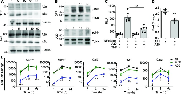
Figure 1. A20 inhibits TNF-induced inflammatory signaling in β cells.
(A and B) Immunoblot of lysates from MIN6 β cells transduced with recombinant adenovirus encoding GFP or human A20 (MOI 100:1) and stimulated with 200 U/mL TNF for the indicated times and probed with antibodies for A20, IĸBα, JNK (T JNK), phospho-JNK (pJNK), or β-actin (loading control). (C and D) βTC3 cells cotransfected with a NF-κB.luciferase reporter (C) or an AP-1.luciferase reporter (D) and a CMV.βgal expression construct ± PCDNA3.1-encoding A20 and stimulated with 200 U/mL TNF for 8 hours or left untreated. RLU, relative light units (luciferase/βgal). (E) Noninfected (NI) MIN6 cells, or GFP- or A20-transduced MIN6 cells, were treated for 1, 4, and 24 hours, and expression of induced genes were assessed. Error bars represent mean ± SEM. Data represent 3 independent experiments, and statistical significance was determined by 1-way ANOVA with Tukey’s multiple comparisons post hoc test (C and E) or 2-tailed Student’s t test (D). *P < 0.05; **P < 0.01; *** P < 0.001.

