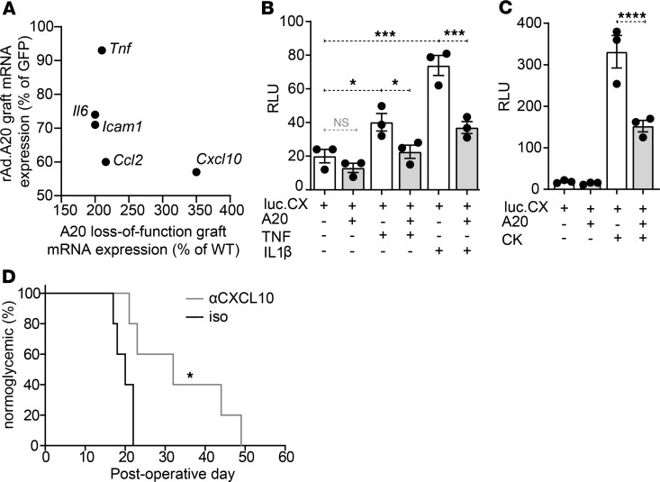
Figure 7. Differential Cxcl10 expression in A20-expressing and loss-of-function islet grafts.
(A) Scatterplot comparing the expression levels (%) of 5 inflammatory genes in allogeneic islet grafts overexpressing A20 (Figure 2H) or harboring loss-of-function A20 (Figure 6H), compared with GFP-expressing control grafts harvested at postoperative day (POD) 10. (B and C) βTC3 cells cotransfected with a CXCL10.luciferase reporter encoding the endogenous promoter (75) and a CMV.βgal-expression construct ± PCDNA3.1-encoding A20. Transfected cells were stimulated with (B) 200 U/mL TNF or IL-1β, or (C) a cocktail (CK) of TNF, IL-1β, and IFNγ for 8 hours or left untreated. Error bars ± SEM. Data representative of 3 independent experiments. Statistical significance determined by 1-way ANOVA with Tukey’s multiple comparisons post hoc test. (D) Percent of C57BL/6 mice normoglycemic after receiving allogeneic BALB/c islet grafts and administered 2 mg/kg of an anti-CXCL10 mAb (n = 5) or an isotype control (iso) (n = 5) by tail vein injection on the day of transplantation and every 2 days thereafter. Significance determined by log-rank test, * P < 0.05; ***P < 0.001; ****P < 0.0001.

