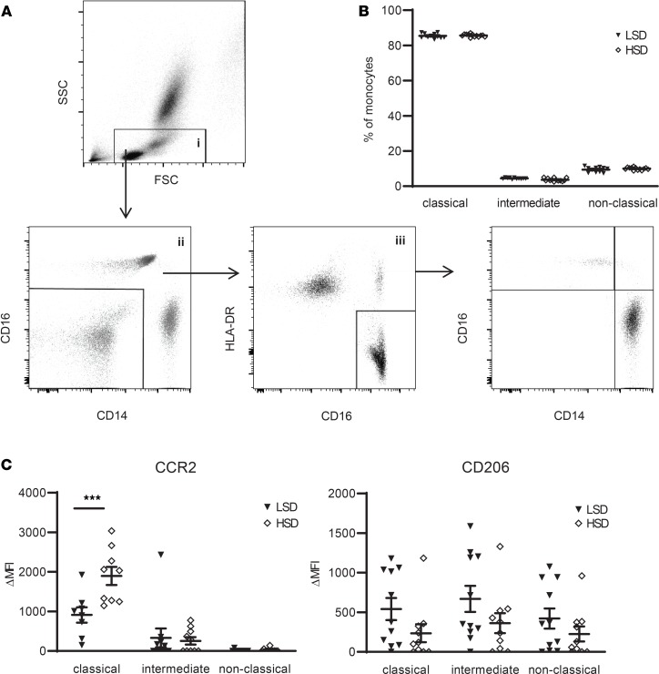
Figure 2. High-salt diet (HSD) induces a proinflammatory monocyte phenotype.
(A) Gating of monocytes and monocyte subtypes. Debris, residual RBC, and granulocytes were first gated out on a FSC/SSC plot (gate i). Gate i was next displayed on a CD14/CD16 plot to select CD14+ and/or CD16+ cells (gate ii). Cells from gate ii were viewed on a CD16/HLA-DR plot to select monocytes (gate iii). This monocyte population was plotted again on a CD14/CD16 plot to gate CD14++/CD16– (classical), CD14++/CD16+ (intermediate), and CD14+/CD16++ (nonclassical) monocyte subsets. (B) Monocyte subsets assessed as described in A, here expressed as a percentage of total monocytes (gate iii) after LSD and HSD. (C) The expression of various surface markers on the monocyte subsets was calculated as ΔMFI = (MFI)positive staining – (MFI)isotype control. CCR2 and CD206 are displayed here; other surface markers are depicted in Supplemental Figure 2. FSC, forward-scattered light. SSC, side-scattered light; LSD, low salt diet; MFI, median fluorescence intensity. Values represent mean ± SEM of n = 11 healthy male volunteers. Data tested using 2-way ANOVA. ***P < 0.001.

