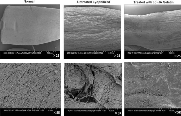
Fig. 6.
Selected scanning electron microscopic images, made after 1000 cycles of tendon excursion, showing normal flexor digitorum profundus tendon, untreated lyophilized tendon, and tendon treated with cd-HA-gelatin. The top row of images was made at low magnification (×25), and the bottom row of images was made at high magnification (×5000). The surface of the lyophilized tendons appeared to be rough, whereas the surface of the lyophilized tendon that had been treated with cd-HA-gelatin appeared to be smoother.

