Fig. 4.
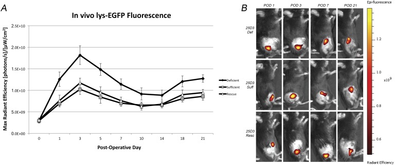
Figs. 4-A and 4-B In vivo neutrophil EGFP fluorescence induced by the Xen36 Staphylococcus aureus strain in 25D3-deficient (def), sufficient (suff), and rescued (resc) mice. A stainless steel implant was inserted into the right knee joint of the mice (n = 20 per group), and the joint space was inoculated with Xen36 S. aureus (1 × 10³ colony-forming units) possessing the bioluminescent construct in a stable plasmid. Fig. 4-A Neutrophil infiltration (neutrophil EGFP fluorescence) as measured by fluorescence in vivo (mean maximum radiant efficiency and standard error of the mean). Fig. 4-B Representative in vivo neutrophil EGFP fluorescence on a color scale overlaid on a grayscale image of the mouse. POD = postoperative day.

