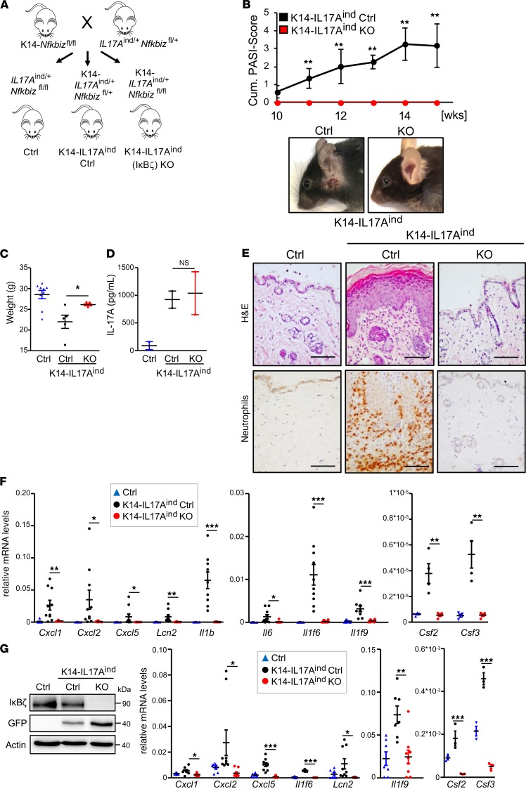
Figure 5. KO of IκBζ in keratinocytes protects against IL-17A–mediated psoriasis.
All analyses (panels C–G) were performed with 15-week-old male mice. (A) Breeding scheme for the generation of keratinocyte-specific Il17a-overexpressing mice with either heterozygous (K14-IL17Aind Ctrl) or homozygous (K14-IL17Aind–KO) deletion of Nfkbiz. (B) Disease progression in K14-IL17Aind mice in the presence or absence of IκBζ. Shown is the cumulative Psoriasis Area and Severity Index (PASI) score accounting for skin lesions, erythema, scaling, and the percentage of the affected skin area. 0 = no phenotype, healthy. 5 = severely affected. n = 6. Bottom: pictures of mice. (C) Body weight of control and IL17Aind mice in the presence and absence of IκBζ. n = 5–9 mice per group ± SEM. (D) Serum levels of IL-17A. n = 2. (E) H&E staining of the back skin and IHC detection of MPO as a marker for neutrophil infiltration. Scale bar: 100 μm. (F) Gene expression of psoriasis-related genes in the skin from 4 to 6 animals per group (mean ± SEM). Depicted are relative mRNA levels normalized to Actin. (G) Left: Immunoblot control of Il17a overexpression and IκBζ deletion in lysates from isolated mKCs. Because the Il17a expression cassette is linked to GFP, expression of IL-17A was monitored by detection of GFP. Actin controlled equal loading. Right: Gene expression analysis in mKCs, isolated from the tails of adult mice. n = 3–9, ± SEM. Relative mRNA levels were normalized to Actin. P values were calculated using 2-tailed Student’s t test (*P < 0.05, **P < 0.01, and ***P < 0.001).

