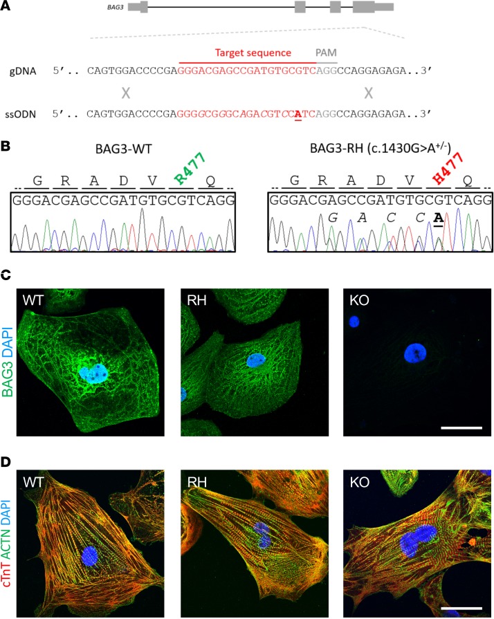
Figure 1. Production and preliminary analysis of BAG3-R477H and BAG3-KO induced pluripotent stem cell (iPSC)-derived cardiomyocytes.
(A) Schematic representation of the BAG3 gene, WT genomic DNA (gDNA) sequence, and the central part of the single-stranded oligonucleotide (ssODN) sequence used to introduce the c.1430G>A (R477H) mutation, which causes DCM in humans (3), into iPSCs. (B) Sanger sequence traces and corresponding amino acid sequences of an unedited iPSC line (BAG3-WT, left) and an iPSC line heterozygous for the c.1430G>A (BAG3-RH) mutation (right). In A and B, underlined/bolded and italicized nucleotides denote the variant of interest and synonymous Cas9-blocking mutations, respectively. (C) BAG3 localization in BAG3-WT (WT), BAG3-R477H (RH), and BAG3-KO (KO) iPSC–derived cardiomyocytes. Green, BAG3; blue, DAPI. Scale bar: 20 μm. (D) Visualization of myofibrillar organization in BAG3-WT (WT), BAG3-R477H (RH), and BAG3-KO (KO) iPSC–derived cardiomyocytes. Red, cardiac troponin T; green, α-actinin; blue, DAPI. Scale bar: 20 μm. Data are representative of 3 independent experiments.

