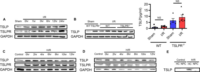
Figure 1. TSLP and TSLPR protein expression increase after liver I/R injury in vivo and H/R in vitro.
(A) TSLP and TSLPR protein expression in liver from WT mice in sham surgery (Sham) or ischemia/reperfusion (I/R; ischemia for 1 hour, reperfusion for 0, 1, 3, 6, 12, or 24 hours) groups were assessed with Western blot. GAPDH served as a loading control. (B) TSLP protein expression of WT and TSLPR–/– mice was assessed in liver (by Western blot) and serum (by ELISA, right) after liver I/R injury (I: 1 hour; R: 6 hours). All data are shown as the mean ± SEM. n = 5 in sham groups, n = 6 in liver I/R groups. NS, no significance. (C and D) TSLP and TSLPR protein expression in primary WT hepatocytes (C) and nonparenchymal cells (D) subjected to hypoxia for 10 hours (1% oxygen) and then reoxygenation for different time points (0, 2, 4, 6, 8, 10, and 12 hours) (H/R). (E) Primary WT hepatocytes (HC) and nonparenchymal cells (NPC) were cultured either in normal oxygen (control group) or in hypoxia for 10 hours (1% oxygen) and then reoxygenation for 8 hours (H/R group). TSLP protein levels in supernatant were assessed with Western blot. For Western blot results, figures are representative of data from multiple mice per experimental group or 3 independent in vitro experiments. ELISA data were assessed by unpaired, 2-tailed Student’s t test (B).

