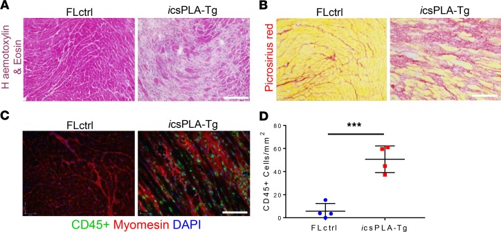
Figure 11. Induction of prelamin A in mouse hearts induces cardiac remodeling and inflammation.
(A) H&E and (B) Picrosirius red staining showed myocardial disarray and fibrosis, respectively. (C) Immunoflourescence micrographs showing CD45+ cells were evident in icsPLA-Tg myocardium. Scale bars: 30 μm. (D) Quantification of CD45+ cells showing an increase in leukocyte population in icsPLA-Tg myocardium. Values are mean ± SD. n = 4 males/group. Unpaired student’s t test was performed. ***P < 0.001.

