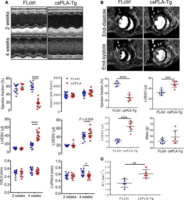
Figure 3. Cardiac function was attenuated in 4-week-old csPLA-Tg mice.
(A) Representative images of echocardiographs and corresponding graphs of analysis performed on movies acquired in B-mode showing severely compromised cardiac function in 4-week-old mice (n = 12/group [6 females, 6 males], except csPLA-Tg 2 weeks, which was n = 9 [7 females, 2 males]). Values are mean ± SD. Two-way ANOVA, no repeated measures, was performed with Sidak’s post hoc test for multiple comparisons. *P < 0.05, ****P < 0.0001. (B) Representative cardiac MRI images of myocardium in end-systole and end-diastole and corresponding graphs displaying a decrease in ejection fraction alongside increases in left ventricle end-diastolic (LVEDV) and LV end-systolic volume (LVESV). Mass was statistically unchanged, but with increased variation, and concurs with postmortem heart weight measurements. (C) Increased relaxation time (R1) of gadolinium contrast in 4-week-old csPLA-Tg myocardium, indicative of fibrosis remodeling. n = 6 males/group. Values are mean ± SD. Student’s 2-tailed t test was performed. **P < 0.01, ***P < 0.001, ****P < 0.0001. IVS;d, intraventricular septal thickness in diastole; LVPW;d, left ventricle posterior wall thickness in diastole.

