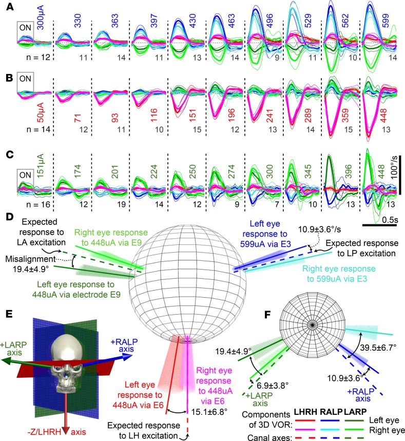
Figure 3. MVI stimulation evokes 3D VOR responses align with targeted semicircular canal.
Mean ± SD cycle-averaged binocular 3D VOR eye velocity responses of subject MVI002 during n cycles of 2 Hz, 40% duty cycle, square-wave-modulated (200 pulses/s for 200 ms, 300 ms off), biphasic, charge-balanced 100 μs/phase current pulse trains. (A) Stimulation via electrode E3 in LP canal ampulla with pulses of 300–599 μA. (B) Stimulation via electrode E6 in LH canal with pulses of 50–448 μA produces a rightward slow phase eye velocity (negative by convention). (C) Stimulation via electrode E9 in LA canal with pulses of 151–448 μA. Right eye response for 396 μA stimulus is missing due to video-oculography tracking failure. (D) Dashed lines denote anatomic semicircular canal axes. Solid vectors depict mean rotation axis for each eye during peak excitatory slow phase response eye velocity for electrodes and currents in adjacent legends. Conic sections denote VOR variability via eigenvalue decomposition of the 3D angular velocity covariance matrix. (E) Canal axes relative to skull landmarks. (F) Same data as D but viewed from above (i.e., from +z/LHRH axis).

