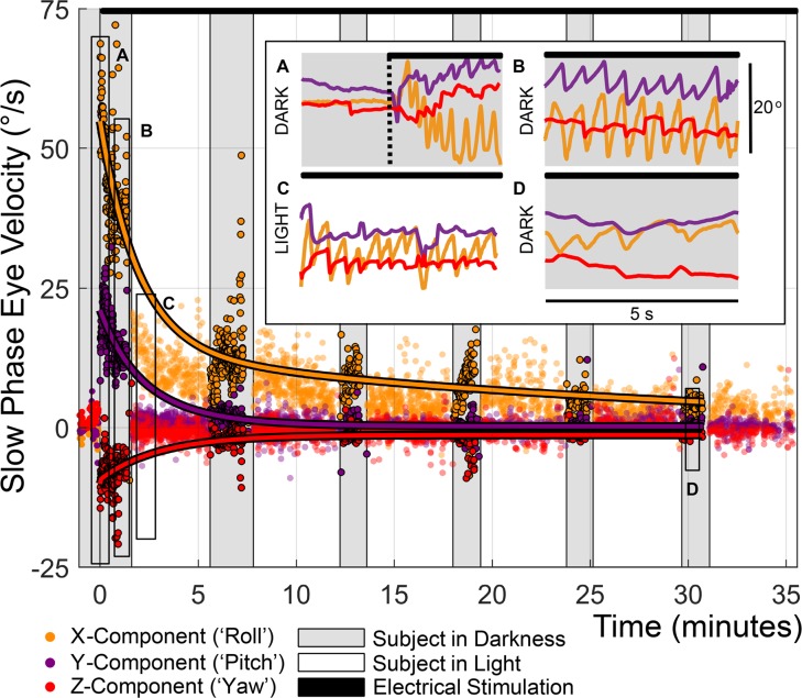
Figure 5. Initial MVI activation elicited robust nystagmus that decayed within 30 minutes.
Three weeks after implantation, continuous electrical stimulation was initiated in subject MVI002 using at 100 pps 100 μs/phase biphasic, charge-balanced current pulses on electrodes in the LP (E3, 599 μA), horizontal (E6, 151 μA) and anterior (E9, 599 μA) canals (black bar at top of figure). Eye movements were monitored for 5-minute cycles of about 1 minute in darkness, then about 4 minutes in light with an Earth-fixed visible target for more than 35 minutes. Each point represents 1 slow phase nystagmus segment. Second-order exponential fits to left eye slow phase velocity in darkness produced dominant time constant estimates of 28.2, 3.33, and 3.0 minutes for roll, pitch, and yaw components, respectively (RMSEx = 6.6°/s, RMSEy = 2.7°/s, and RMSEz = 2.7°/s). Insets show (A) robust nystagmus dominated by positive slow phase torsional component at onset of stimulation, (B) extinction of the horizontal nystagmus component in darkness by t = 1 minutes, (C) suppression of all but the ~18°/s torsional response during lights-on testing, and (D) a reduction of the torsional component to ~5°/s by t = 30 minutes.

