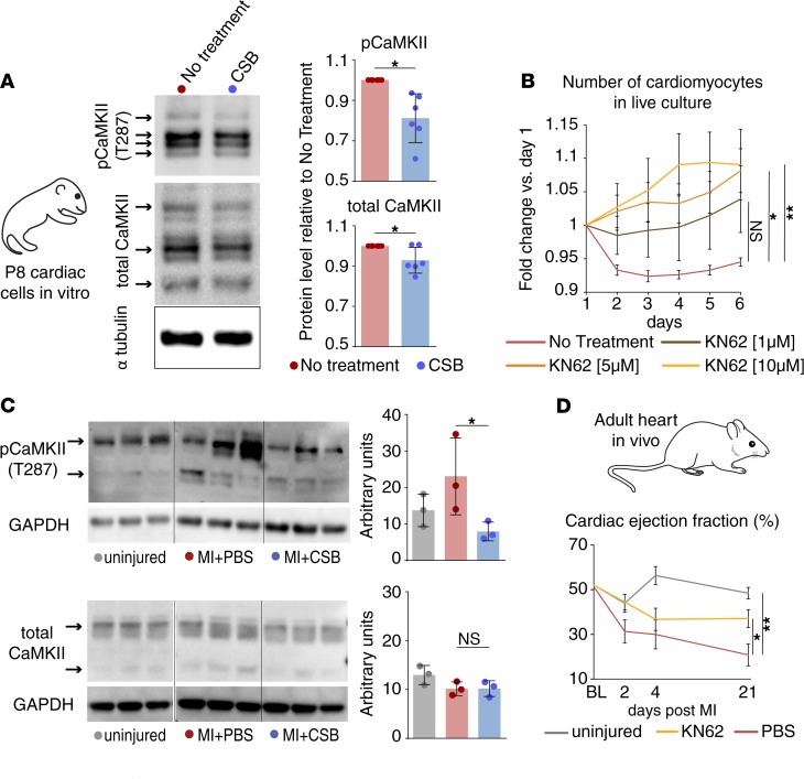
Figure 4. CaMKII inhibition by CSB improves heart function after MI.
(A) Western blot images and quantification show the protein level of total CaMKII and phospho-CaMKII (pCaMKII) in P8 cardiac cell cultures treated with CSB for 4 days versus untreated control (n = 6 for each group, mean ± SD, paired 2-tailed Student’s t test). (B) Number of P8 cardiomyocytes in live culture relative to day 1 as quantified by the screening system (no treatment, n = 11; 1 μM KN62, n = 4; 5 μM KN62, n = 7; 10 μM KN62, n = 7; mean ± SEM, 1-way ANOVA and Dunnett’s post hoc test). (C) Western blot images and quantifications show the protein level of total CaMKII (lower panel) and pCaMKII (upper panel) in the ventricles of adult mice 5 days after MI (n = 3 for each group, mean ± SD, 1-way ANOVA and Sidak’s correction). Samples from uninjured mice were run in the same gel as MI + PBS and MI + CSB but were noncontiguous. (D) Cardiac EF of adult mice uninjured (n = 3) or injured and treated with PBS (n = 6) or KN62 (n = 6) up to 21 days after MI (mean ± SEM, 1-way ANOVA and Dunnett’s post hoc test). For all panels: *P < 0.05, **P < 0.01. NS, not significant.

