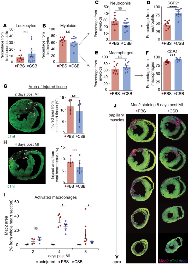
Figure 7. CSB inhibits neutrophil and macrophage activation in the heart following MI.
(A–C and E) FACS analysis shows the percentage of total leukocytes (A), myeloid cells (B), neutrophils (C), and macrophages (E) in the heart of adult mice 4 days after MI. (D and F) Percentage of CCR2+ neutrophils (D) and macrophages (F) in the hearts of adult mice 4 days after MI. Results for A–F were obtained from 2 separate experiments. PBS, n = 9; CSB, n = 10; data presented as mean ± SEM, unpaired 2-tailed Student’s t test. (G–J) Mac2 staining in injured hearts. Adult mice underwent MI and were treated with PBS or CSB in the same manner as in the ELISA experiment (1 i.m. and 3 i.v. injections). Hearts were isolated 2, 4, and 8 days after MI and stained for cardiac troponin I (cTnI) as a cardiomyocyte marker and Mac2. (G) Representative image of injured heart 2 days after MI and quantification of the injured zone size in PBS-treated (n = 4) or CSB-treated (n = 5) mice (mean ± SD). (H) Representative image of injured heart 4 days after MI and quantification of the injured zone size in PBS-treated (n = 5) or CSB-treated (n = 6) mice (mean ± SD). (I) Quantification of activated macrophages in the hearts of adult mice 2 days (uninjured, n = 4; PBS, n = 4; CSB, n = 5), 4 days (uninjured, n = 3; PBS, n = 5; CSB, n = 6), and 8 days (uninjured, n = 3; PBS, n = 7; CSB, n = 8) after MI. Quantification was performed by measuring the area stained for Mac2 as percentage of the area of the whole section (mean ± SEM). (J) Representative serial sections of adult mouse hearts stained for Mac2 8 days after MI. Statistical analysis for G–I was performed using 1-tailed, unpaired Student’s t test. For all panels: *P < 0.05, ***P < 0.001, ****P < 0.0001. NS, not significant.

