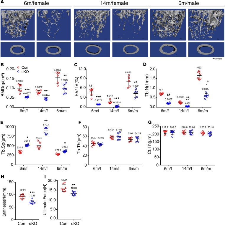
Figure 2. Pinch loss causes severe osteopenia in 6- and 14-month-old male and female mice.
(A) Three-dimensional (3-D) reconstruction from micro-computerized tomography (μCT) scans of distal femurs from 6- and 14-month-old female and 6-month-old male control and dKO mice. Scale bar: 100 μm. (B–G) Quantitative analyses of bone mineral density (BMD), bone volume/tissue volume (BV/TV), trabecular number (Tb.N), trabecular thickness (Tb.Th), trabecular separation (Tb.Sp), and cortical thickness (Ct.Th) of distal femurs from A. n = 6 mice per group. *P < 0.05, **P < 0.01, ***P < 0.001 vs. controls. Unpaired Student’s t test. Results are expressed as mean ± SD. (H and I) Three-point bending tests. Femurs isolated from 4-month-old control and dKO female mice were subjected to the 3-point bending test as described in Materials. Stiffness (H) and maximum load (I). n = 8 mice per group. **P < 0.01, ***P < 0.001 vs. controls. Unpaired Student’s t test. Results are expressed as mean ± SD.

