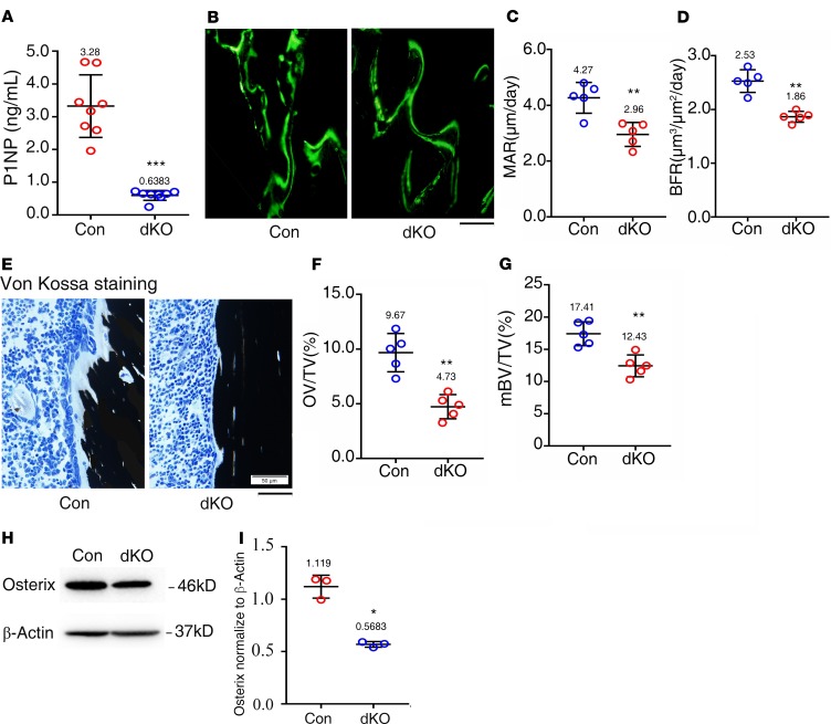
Figure 3. Pinch loss impairs osteoblast formation and function in bone.
(A) Serum level of procollagen type 1 amino-terminal propeptide (P1NP). Sera were collected from 6-month-old female mice of the 2 genotypes. n = 8 mice per group. ***P < 0.001 vs. controls. Unpaired Student’s t test. Results are expressed as mean ± SD. (B–D) Calcein double labeling. Representative images of 3-month-old female control and dKO tibial sections (B). Sections of nondemineralized tibiae of 3-month-old female control and dKO mice were used for measurements of mineralization apposition rate (MAR)of the metaphyseal trabecular bones (C) and bone formation rate (BFR) (D). n = 5 mice per group. **P < 0.01 vs. controls. Unpaired Student’s t test. Results are expressed as mean ± SD. Scale bar: 50 μm. (E–G) von Kossa staining. Undecalcified sections of femora from 3-month-old female control and dKO mice were subjected to von Kossa staining (E). Quantitative osteoid volume/tissue volume (OV/TV) (F) and mineralized bone volume/tissue volume (mBV/TV) (G) data for the cancellous bones from distal femora were measured by bone morphometry. n = 5 mice per group. **P < 0.01 vs. controls. Unpaired Student’s t test. Results are expressed as mean ± SD. Scale bar: 50 μm. (H) Western blot analyses. Protein extracts were isolated from femurs of 6-month-old female control and dKO mice and subjected to Western blotting using an antibody against osterix. (I) Quantitative data. *P < 0.05 vs. controls. Unpaired Student’s t test. Results are expressed as mean ± SD. Western blotting was repeated 3 times.

