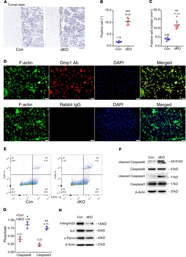
Figure 7. Reduced integrin β3, ILK, and α-parvin; increased active caspase-3 and -8 expression; and apoptosis in Pinch-deficient osteocytes.
(A–C) TUNEL staining. Tibial sections of 6-month-old female control and dKO mice were subjected to TUNEL staining (A), percent apoptotic osteocytes (B), and the number of osteocytes (C) in the cortical bone matrix were quantified. n = 7 mice per group. **P < 0.01, ***P < 0.001 vs. controls. Unpaired Student’s t test. Results are expressed as mean ± SD. (D) Primary osteocytes were isolated from middiaphyseal femoral shafts (with their BM flushed) of 9-month-old female control and dKO mice and subjected to immunofluorescence (IF) staining with rabbit DMP1 antibody, normal IgG, or Alexa Fluor 488 Phalloidin (for F-actin) (D). (E) Primary osteocytes from D were subjected to flow cytometry analysis of cell apoptosis. Scale bar: 20 μm. (F and H) Western blot analysis. Protein extracts were isolated from middiaphyseal femoral shafts (with their BM flushed) of 6-month-old female control, and dKO mice were subjected to Western blotting analyses using the indicated antibodies. (G) Quantitative data of F. n = 3. *P < 0.05, **P < 0.01, versus controls, unpaired Student’s t test. Results are expressed as mean ± SD. Western blotting analyses in this figure were repeated at least 3 times.

