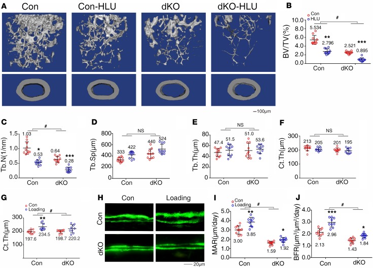
Figure 8. Pinch deletion exacerbates hindlimb unloading-induced bone loss and reduces active ulna loading–stimulated bone formation.
(A–F) Hindlimb unloading (HLU) experiment. Three-month-old female control and dKO mice were treated with or without tail suspension for 21 days, followed by μCT analysis of femurs. (A) Three-dimensional (3-D) reconstruction. Scale bar: 100 μm. (B–F) Quantitative analyses of BV/TV, Tb.N, Tb.Sp, Tb.Th, and Ct.Th. n = 10 mice per group. *P < 0.05, **P < 0.01, ***P < 0.01, control (Con) vs. HLU; #P < 0.05, Con-HLU/Con vs. dKO-HLU/dKO, 2-way ANOVA. Results are expressed as mean ± SD. (G–J) Ulna loading experiment. Three-month-old female control and dKO mice were subjected to ulna loading as described in Methods. Quantitative analyses for Ct.Th (G). Sections of nondemineralized femurs of control and dKO mice with and without loading were used for measurements of mineralization apposition rate (MAR) and bone formation rate (BFR) (H–J). Quantitative MAR and BFR data for diaphyseal cortical bones. n = 9 mice per group. *P < 0.05, **P < 0.01, ***P < 0.01, Con vs. load; #P < 0.05, Con-load/Con vs. dKO-load/dKO, 2-way ANOVA. Results are expressed as mean ± SD. Scale bar: 20 μm.

