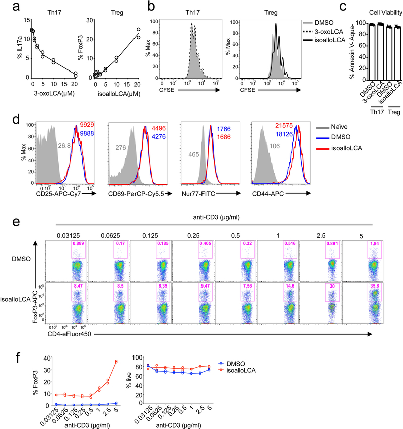
Extended Data Figure 4. Effects of isoalloLCA on FoxP3 expression require strong TCR stimulation.
a, 3-oxoLCA and isoalloLCA demonstrate dose-dependent effects on Th17 cell and Treg differentiation, respectively (n = 2). A low concentration of TGF-β (0.01 ng/ml) was used for Treg culture. b–d, 3-oxoLCA and isoalloLCA do not significantly affect cell proliferation, cell viability, or T cell activation. b, Naïve CD4+ T cells were labeled with a cell proliferation dye CFSE and cultured for 3 days in the presence of DMSO, 3-oxoLCA or isoalloLCA under Th17 or Treg polarization conditions. c, Live cell percentages at the end of the 3-day culture were determined based on both Annexin V and fixable live/dead staining (n = 3/group). d, Both DMSO and isoalloLCA treatment lead to comparable levels of expression of CD25, CD69, Nur77 and CD44. Naïve CD4+ T cells were used as a negative control. e and f, T cells were cultured with different concentrations of anti-CD3 antibody, in the presence of DMSO or isoalloLCA (20 μM). Representative FACS plots of CD4+ T cells cultured for 3 days and stained intracellularly for FoxP3 (e). Quantification of FoxP3+ and viable T cells after 3-day culture (f) (n = 2/group). n, number of biologically independent samples. Data are representative of two independent experiments (b, d). Data in (c) are shown as the mean ± standard deviation.

