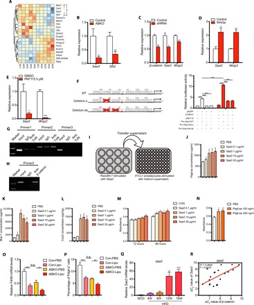
Fig. 6. β-catenin ablation in mature adipocyte inhibits preadipocyte proliferation partially mediated by repressing Saa3 and decreasing macrophage recruitment.

(A) Gene expression profiles of iWAT in 24-week HFD-fed control and ABKO mice. Heatmap of genes that are differentially expressed between two groups, with a fold change larger than 1.5 and the corresponding P values less than 0.05. (B) Validation of Saa3 expression in iWATs between the two groups by real-time polymerase chain reaction (PCR). (C) Saa3 expression detected in mature adipocytes differentiated from 3T3-L1 cells after β-catenin knockdown by shRNA. (D) Saa3 expression detected in mature adipocytes differentiated from 3T3-L1 cells after canonical Wnt signaling activation by Wnt3a (10 ng/ml) treatment for 16 hours. (E) Saa3 expression detected in mature adipocytes differentiated from SVF cells after canonical Wnt signaling inhibition by 5 μM PKF115 treatment for 48 hours. (F) Left: Schematic of wild-type Saa3 promoter and mutations deleting the indicated Tcf4 binding sites. Right: Luciferase reporter assay performed in the 3T3-L1 cell line 48 hours after transfection with the indicated plasmids. pRL-TK (expressing Renilla luciferase) was used as the normalized control. (G) Chromatin immunoprecipitation (ChIP) assay in white adipocytes differentiated from SVF immunoprecipitated with anti-Tcf4 antibody followed by PCR detection with the indicated primers for different parts of Saa3 promoter. Of note, products with primer 2 were designed to amplify the region containing Tcf4 binding sites, while primer 1 and primer 3 were not. IgG, immunoglobulin G. (H) ChIP assay in differentiated white adipocytes immunoprecipitated with anti–β-catenin antibody followed by PCR detection with the primer 2. (I) Schematic of experiment design in which Raw264.7 was stimulated with recombinant Saa3 for 6 hours, and the supernatant was then added to 3T3-L1 cells and refreshed every 24 hours. (J to L) Concentrations of Pdgf-aa (J), Mcp-1 (K), and Cxcl2 (L) in the conditioned medium of Raw264.7 cells treated with recombinant Saa3 at 0.1, 1, 10, and 30 μg/ml, respectively. (M) Viability of 3T3-L1 cells treated with the conditioned medium of Raw264.7 cells in (I). (N) Cell viability of 3T3-L1 cells treated with the recombinant Pdgf-aa protein with different concentrations for 120 hours. (O and P) The relative mRNA levels of F4/80 (O) and the percentage of Pdgfrα+ cells calculated (P) in the indicated groups (n = 6 to 8 for each group). (Q) Saa3 expression was induced in iWAT during long-term HFD feeding (n = 4 for each group). (R) Saa3 expression was associated with β-catenin expression in iWAT. ΔCt value of Saa3 and β-catenin were used to examine the association in the linear regression analysis. ABKO, Adiponectin-cre;β-cateninflox/flox; HFD, high-fat diet; Con-PBS, control mice injected with PBS; Con-Lipo, control mice injected with liposome; ABKO-PBS, ABKO mice injected with PBS; ABKO-Lipo, ABKO mice injected with liposome. Data are shown as means ± SEM. *P < 0.05, **P < 0.01, ***P < 0.001. n.s., not significant.
