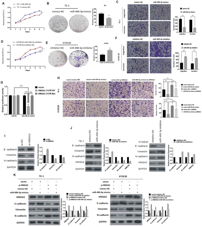
Figure 5.
The function of miR-490-3p in ESCC cells. (A) and (B) The effect of miR-490-3p on cell proliferation in vitro using CCK8 assay and colony formation assay after overexpressing miR-490-3p in TE-1. (C) Cell migration and invasion abilities were assessed by transwell assay after overexpressing miR-490-3p in TE-1 cells. (D and E) The effect of miR-490-3p on cell proliferation in vitro using CCK8 assay and colony formation assay after knocking down miR-490-3p in KYSE30 cells. (F) Cell migration and invasion abilities were assessed by transwell assay after knocking down miR-490-3p in KYSE30 cells. (G) Luciferase reporter assay for the luciferase activity of HMGA2-3 ′UTR WT or HMGA2-3 ’UTR mutant in cells cotransfected with miRNA mimics. (H) The invasion ability was evaluated by transwell Matrigel invasion assays (I) Knockdown of HMGA2 inhibits EMT (J) Western blot analysis comparing upregulated and downregulated- miR-490-3p ESCC cells with control cells were shown for vimentin, E-cadherin, N-cadherin and HMGA2. (K) The expression of HMGA2 and EMT markers was detected by Western blot after transfection with mimics or HMGA2 overexpression plasmids.* P<0.05,**P<0.01, **P<0.001.

