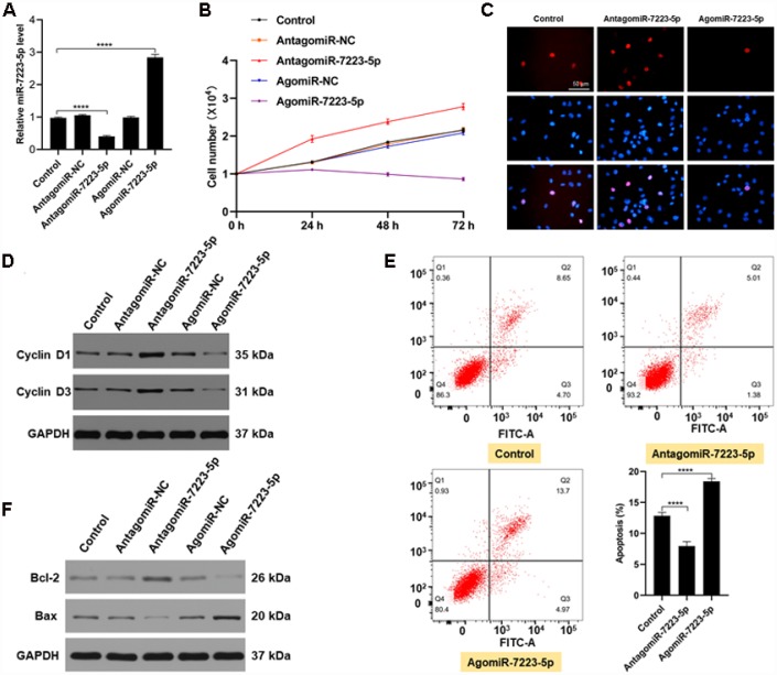
Figure 2.
miR-7223-5p suppressed the function of MC3T3-E1 cells in vitro. (A) miR-7223-5p levels in MC3T3-E1 cells were measured by qRT-PCR after transfection with PBS (control), antagomiR-NC, antagomiR-7223-5p, agomiR-NC, or agomiR-7223-5p. (B, C) CCK8 and Edu assays were used to assess the proliferation of MC3T3-E1 cells after transfection with agomiR-7223-5p and antagomiR-7223-5p. (D) Cyclin D1 and cyclin D3 were detected by western blot after transfecting cells with agomiR-7223-5p or antagomiR-7223-5p. (E) The percentage of apoptotic MC3T3-E1 cells (Q2+Q3) was measured by flow cytometry 24 h after transfection with miR-7223-5p. Q1: dead cell; Q2: later apoptosis; Q3: early apoptosis; Q4: living cells. (F) Western blot showing Bax and Bcl-2 levels after transfecting cells with agomiR-7223-5p and antagomiR-7223-5p.

