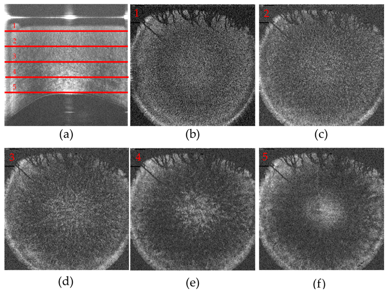
Figure 5.
Volume data of the cornea recorded in the cross-polarized detection channel. (a) Representative B-scan (lateral average of 10 frames) The depth location of the corresponding en-face images is marked with red horizontal lines; (b)–(f) En-face frames retrieved at a depth of 100, 200, 300, 400, 500 pixels (101, 202, 303, 404, 505 μm) from the corneal surface (cf. red lines 1–5 in (a); The field of view of (a) is 11 mm (x) and 860 μm (z), of Figure 5b–f is 11 mm (x) and 11 mm (y).

