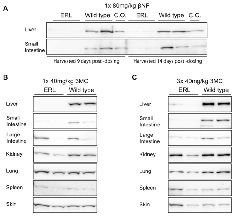
Figure 2. Deletion of P450 oxidoreductase with β-NF or 3-MC in various tissues.
(A) Western blots of POR expression in the liver and small intestine of ERL (Porlox/lox::CreCyp1a1 − KI) and WT (Porlox/lox) mice (n = 2) given a single injection of βNF (80 mg/kg i.p.). Tissues were harvested either 9 or 14 days later and individual microsomal fractions blotted as described. WT mice treated with corn oil (C.O.) only were used as vehicle control. (B) Western blots of POR expression in the liver, small intestine, lung, kidney, spleen and skin of ERL (Porlox/lox/CreCyp1a1 − KI) and WT (Porlox/lox) mice (n = 2) dosed with a single injection of 3MC (40 mg/kg i.p.). Tissues were harvested 14 days later, and individual microsomal fractions blotted as described. (C) Western blots of POR expression in the liver, small intestine, lung, kidney, spleen and skin of ERL (Porlox/lox::CreCyp1a1 − KI) and WT (Porlox/lox) mice (n = 2) dosed with 3MC (40 mg/kg i.p.) on three consecutive days. Tissues were harvested 14 days after the initial dose and individual microsomal fractions blotted as described.

