Fig. 1.
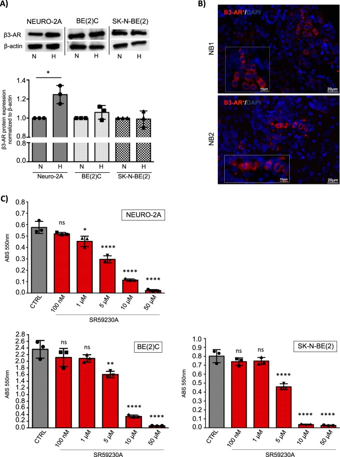
β3-AR is constitutively expressed in murine and human neuroblastoma cells and it is involved in cell survival. a WB and relative densitometric quantification analysis, showing expression of β3-AR protein in murine (Neuro-2A) and human (BE(2)C, SK-N-BE(2)) NB cell lines in normoxic (N) and hypoxic condition (H). Results were normalized to the expression of β-actin and reported as mean ± SD, fold change over controls (normoxic condition), set as 1. Blots are representative of three independent experiments. Significance was calculated by Unpaired t-test analysis with equal SD (*P < 0.05). b Immunofluorescence staining of β3-AR on tumor sections of two NBs (NB1, NB2) derived from human patients, showing numerous β3-AR positive cells. Images are representative of similar results obtained for n = 6. c MTT survival assay in Neuro-2A, BE(2)C and SK-N-BE(2) NB cell lines, treated with different concentration of SR59230A for 24 h. Results are reported as mean ± SD of three independent experiments performed in triplicate. Significance was calculated by one-way ANOVA analysis followed by Bonferroni’s post hoc test (ns = not significant, *P < 0.05, **P < 0.01, ****P < 0.0001)

